
Process of photosynthesis
- Photosynthesis is a chemical process that uses sunlight to turn carbon dioxide into sugars the cell can use as energy.
- Carbon dioxide + Water + light energy --> Glucose + Oxygen
- Photosynthesis has two main sets of reactions. Light-dependent reactions need light to work; and light-independent reactions, which do not need light to work.
Source and function of raw materials of photosynthesis
The sources and functions of raw materials of photosynthesis are as follows:
Carbon dioxide
Carbon dioxide
- It is the source of carbon that required for the synthesis of sugar.
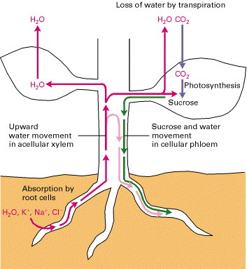
- It is obtained from the atmosphere. It enters the leaf by diffusion through stomata.
- Apart from carbon, hydrogen is also needed to prepare sugar. Water is the source of hydrogen ion.
- From the soil water is taken up by the roots and sent to the leaves through xylem tissues.
- Due to other metabolic process and transpiration, an adequate supply of water is always needed.
- Light is needed for the light-dependent stage of photosynthesis.
- Most of the plants use sunlight as a source of energy. But some used artificial light e.g., plantlets produced by tissue culture.
- If plant lack light for a substantial period of time they will die due because stored starch have been used up by them.
Process of photosynthesis
The process of photosynthesis occurs in the mesophyll cells because it contains numerous chloroplasts.
- Chloroplasts contain chlorophyll pigment that absorbs sunlight and provides energy.
- The lower surface of leaves has numerous pores called as stomata. It diffuses the carbon dioxide from the air into the leaves.
- In this process, water and carbon dioxide prepares glucose (food) and releases oxygen.

Importance of photosynthesis
The process of photosynthesis plays an important role in such ways:
- Plant prepares their food by photosynthesis. The plants, in turn, are eaten by the animals.
- Photosynthesis convert radiant or solar energy into chemical energy.
- Productivity of agricultural crops directly depends upon the rate of photosynthesis.
- It provides oxygen in atmosphere for all living organisms.
- It maintains the balanced level of oxygen and carbon dioxide ecosystem.
- Fossil fuels are derived from plants. Energy stored in fuel is originally trapped from sun druring photosynthesis.
Mechanism of water absorption
It is of two types, active and passive.
- Passive water absorption is the type of water absorption which originates in the aerial parts of the plants due to loss of water in transpiration.
- Active water absorption is the absorption of water due to forces present in the root.
Transportation of food and other substances
- Excess of food is taken into the storage organs like roots, fruits and seeds.
- This process is known as translocation and it takes place through phloem in upward as well as downward direction.
- In flowering season, sugar stored in the roots or stem is translocated to the buds for growing them into flowers.
Heart

The heart
is the central organ for pumping the blood throughout the body. Heart
is made up of strong cardiac muscles. It is located in the chest cavity
with its lower part pointing towards the left. Its size is that of the
persons fist. It pumps blood rich in carbon dioxide to the lungs and
oxygen-rich blood to other parts of the body.
- The heart
consists of four chambers namely auricles and ventricles. The two upper
chambers of the heart are known as the auricles. - The two
lower chambers of the heart are the ventricles. Left and right parts of
the heart are separated by a muscular partition called as septum. - Heart has
number of valves which allow the blood to flow in one direction. These
prevent the oxygenated blood mixing with de-oxygenated blood. - SA node, the natural and primary pace maker of the heart is located in the upper wall of the right atrium in the heart.
- AV node,
the secondary pace maker is located in the bundles of tissues on the
border between right atrium and right ventricle of the heart.
Valves in the heart
Valves in the heart regulate the unidirectional flow of blood.
- Bicuspid between the left atrium and the left ventricle.
- Tricuspid between the right atrium and the right ventricle.
- Semilunar valve between ventricles and aorta and between ventricles and pulmonary artery.
- Sharp closing of these valves produces the Lubb and Dup sounds.
General features of Veins
Veins
- It carries blood from organs to the heart.
- All the veins carry impure blood except pulmonary vein which carries pure blood.
- In veins, blood flows with a low pressure and speed.
- Veins are superficial placed just below the skin.
- The walls of the artery are thick and tough.
- Their lumen is wide.
General features of Capillaries
Capillaries
- Capillaries are smallest blood vessels.
- The arterioles and venules further divide into thin walled vessels called capillaries.
- The capillaries of arteries and veins are united.
- It helps in the diffusion of nutrients, hormones, gases etc. into the tissue cells.
General features of Artery
Artery
- It carries blood from the heart to the organs.
- All the arteries carry pure blood except pulmonary artery which carries impure blood.
- Blood flows with a high pressure and speed.
- Arteries are deeply situated in the body.
- The walls of the artery are thick and tough.
- Their lumen is constricted.
Characteristics of an artery
The blood vessels that carry blood away from the heart to other parts of the body are called arteries.
- Arteries carry oxygenated blood (except pulmonary artery).
- Thick walled
- Elastic
- Muscular
- Valves absent
- Narrow lumen
- Deeply situated
- Arteries further divide to form arteriole and further divide to form capillaries.
- Wall of artery is made up of tunica externa, tunica media and tunica interna.
Characteristics of vein
The blood vessels that carry blood from various organs of the body to the heart are called veins.
- Veins carry deoxygenated blood (except pulmonary veins)
- They are thin-walled
- They Possess semilunar valves
- They are superficially situated
- They have a broad lumen
- Wall of the vein is made up of tunica externa, tunica media and tunica interna.
Major arteries of Human body.
Major arteries in the human body are
- Aorta - The largest artery in the human body.
- Coronary arteries - supply oxygenated blood to the muscles of the heart.
- Pulmonary arteries - carries deoxygenated blood from the right ventricle to the lungs. It is the only artery that carries deoxygenated blood.
- Common carotid artery - supply oxygenated blood to head and neck.
- Internal carotid artery - supply oxygenated blood to the brain.
- Subclavian artery - supply oxygenated blood to the shoulders and arms.
- Superior mesenteric artery - supply blood to the small intestine.
- Inferior mesenteric artery - supply blood to the large intestine.
- Internal iliac - supply blood to organs of the pelvic region.
- External iliac - supply blood to lower extremities.
- Hepatic artery - supply blood to the liver.
- Renal artery - supply blood to the kidneys.
Major veins in human body
The major veins in human body are
- Vena cava ( superior and inferior) - These are the largest veins. It collects the deoxygenated blood from other veins in upper and lower portion of the body and passes it to the right atrium.
- Saphenous vein - it is the longest vein in the body.
- Pulmonary vein - carry oxygenated blood from the lungs to the heart. It is the only artery that carries deoxygenated blood.
- Hepatic vein - brings back deoxygenated blood from liver towards the heart.
- Renal vein brings blood from the kidney back to the heart.
- External jugular vein - returns blood from the outer cranial, face, and neck towards the heart.
- Internal jugular vein - returns blood from the brain towards the heart.
- Superior mesenteric vein - Collects blood from a section of small intestine. It is one of the veins that flow into the portal vein.
Circulation of blood in the heart

GIF

The circulation of blood within the different chambers of the heart is described below:
Right atrium:
Right atrium:
- Deoxygenated blood enters the right atrium from
- The anterior region of the body via the superior vena cava and the posterior region of the body via the inferior vena cava.
- This causes an expansion of the right atrium.
- When the right atrium contracts, the tricuspid valve is opened.
- As a result, the blood flows from the right atrium into the right ventricle.
- When the right ventricle contracts, the blood is forced into the pulmonary artery due to the opening of the pulmonary semilunar valves. The pulmonary artery carries the blood to the lungs for oxygenation.
- After oxygenation, four pulmonary veins bring the blood back from the lungs into the left atrium.
- This makes the left atrium expand.
- When the left atrium contracts, the bicuspid valve is opened.
- As a result, the blood flows from the left atrium into the left ventricle.
- When the left ventricle contracts, the oxygenated blood is pushed into the aorta via the aortic semilunar valves.
- The aorta then carries the oxygenated blood to different parts of the body.
Double circulation
A type of circulation in which blood flows through the heart twice is called double circulation. This type of circulatory system has separate systemic circulation and pulmonary circulation.
- Systemic circulation - The flow of oxygenated blood from the left ventricle of the heart to various parts of the body and deoxygenated blood from various parts of the body to the right atrium is called systemic circulation. The systemic arteries arising from aorta carry oxygenated blood from the left of the ventricle to various parts of the body. The systemic veins carry deoxygenated blood from various parts of the body to the right atrium of the heart.
- Pulmonary circulation - The flow of deoxygenated blood from the right ventricle to the lungs and the return of oxygenated blood from the lungs to the left atrium is called pulmonary circulation. The pulmonary trunk( right and left pulmonary artery) carries blood from the right ventricle to the lungs where the exchange of gases takes place. The oxygenated blood from the lungs returns to the left atrium of the heart through two pulmonary veins, one from each lung.
Double circulation
- The majority of mammals (including humans) utilize a double
circulatory system. - This means that we have two loops in our body in
which blood circulates. - One is oxygenated, meaning oxygen rich, and the
other is deoxygenated, which means it has little to no oxygen, but a lot
of carbon dioxide.
Photoautotroph
Photoautotrophic mode of nutrition is the process of formation of food by the process of photosynthesis.
- In this process, the organism used sunlight as a source of energy along with carbon dioxide and water.
- An organism that follows such mode of nutrition is called as photoautotroph e.g., algae, green plants.
Autotrophic nutrition
Mode of nutrition is the way to obtain food. On the basis of obtaining and utilizing food, there are two types, autotrophic and heterotrophic.
- Autotrophic nutrition: When an organism prepared their own food and does not depend on any other organism is called as autotrophic nutrition.
- An organism that follows the autotrophic mode of nutrition is called as autotrophs e.g., plants.
- Autotrophic mode of nutrition is further divided into two categories photoautotrophs (uses photosynthesis as a source of energy, e.g., plants and green bacteria) and chemoautotrophs (uses chemosynthesis, e.g., non- green sulphur bacteria).
Hetertrophic nutrition
The mode of nutrition in which an organism depend on the plant or another animal for food is called as heterotrophic nutrition. An organism that carries out heterotrophic nutrition is called as heterotrophs e.g., animals.
Heterotrophic mode of nutrition is further divided into three groups such as:
Heterotrophic mode of nutrition is further divided into three groups such as:
- Holozoic: It is the method in which animal taking in the complex food e.g., human being, cow, lion deer etc.
- Saprophytic: It is the method in which organism first convert the complex food into the simpler and then use them as food e.g., bacteria, fungi etc.
- Parasitic nutrition: It is the method in which an organism lives in the body of another organism and get nutrients from them e.g., Plasmodium, worms etc.
Difference between autotrophic and heterotrophic nutrition
Different organisms obtain food by different modes such as autotrophic and heterotrophic mode of nutrition. These modes of nutrition have some differences which are as follows:
| Autotrophic nutrition | Heterotrophic nutrition |
| 1. Organisms prepare their own food. | Organisms depend on another organism for food. |
| 2. It includes photosynthesis and chemosynthesis. | It includes holozoic, saprotrophic and parasitic. |
| 3. Organisms have chlorophyll pigment. | Organisms do not have chlorophyll. |
| 4. Organisms are called as producers. | Organisms are called as consumers. |
Types of heterotrophic nutrition
Based on the source of nutrition, heterotrophic mode of nutrition can be divided as follows:
- Saprophytic nutrition: In this mode of nutrition, organisms obtain nutrients from dead and decaying organic matter. These organisms are called Saprophytes. Example: Yeast
- Parasitic nutrition: In this mode, the organism lives inside or outside the body of another organism. It takes nutrition from the other organism without killing it. Such organisms are called parasites. Example: Ascaris (roundworm) are parasites on the human body.
- Holozoic nutrition: Organisms which show holozoic nutrition feed on complex matter. This matter is digested and nutrients are absorbed. Example: Human beings
Feeding and digestion in amoeba
- Amoeba is a microscopic organism which has fingerlike known as pseudopodia.
- The amoeba extends its pseudopodia around the food and engulfs it.
- The food is trapped in the food vacuole where it is digested by the digestive enzyme and finally the food is absorbed and distributed all through the body.
Feeding and digestion in Amoeba

Amoeba is a unicellular organism. It undergoes intracellular digestion. The mode of nutrition in Amoeba is holozoic.
The process of obtaining food by Amoeba is called phagocytosis.
Ingestion: When a food particle is near the Amoeba, it forms temporary finger-like projections called pseudopodia around the food particle and engulfs it.
Digestion: The food is digested in the food vacuole with the help of enzymes.
Absorption: It is then absorbed in the cytoplasm of the Amoeba by diffusion.
Assimilation: The absorbed food provides energy and a part of nutrition is used for growth.
Egestion: The undigested and the waste food particles are thrown out.
Parts of the digestive system

The food is broken down into smaller pieces by the teeth and mixed with the saliva in the mouth. Saliva moistens the food and helps in digestion of carbohydrates. With the help of the tongue, the food is pushed forward.
Food particles then move to the stomach through the esophagus. Esophagus contains sphincters to control movement food. In the stomach, the food particles are mixed with hydrochloric acid and other digestive juices. The digested food is called chyme. Chyme is then moved to the small intestine. Digestive juices from pancreas, liver, and gallbladder are mixed for further chemical digestion. The small intestine absorbs nutrients. The chyme is then passed to the large intestine. In the large intestine, water absorption takes place. The undigested part changes to solid stool. The rectum stores the undigested food till the waste is passed through the anus.
Food particles then move to the stomach through the esophagus. Esophagus contains sphincters to control movement food. In the stomach, the food particles are mixed with hydrochloric acid and other digestive juices. The digested food is called chyme. Chyme is then moved to the small intestine. Digestive juices from pancreas, liver, and gallbladder are mixed for further chemical digestion. The small intestine absorbs nutrients. The chyme is then passed to the large intestine. In the large intestine, water absorption takes place. The undigested part changes to solid stool. The rectum stores the undigested food till the waste is passed through the anus.
Carhohydrate digestion in the oral cavity
- The salivary glands in the oral cavity secrete saliva, which helps to moisten the food.
- The food is then chewed while the salivary glands also release the enzyme salivary amylase, which begins the process of breaking down the starch into maltose, isomaltose and alpha dextrins.

Dental formula
Dental formulae indicate the number of each type of tooth in a given species.
The jaw is bilaterally symmetrical. Hence, the dental formulae represent the number considering the half jaw only.
The numerator of the fraction represents the number of teeth in the upper jaw and denominator represents the number of teeth in the lower jaw.
The formula represents number of teeth in the following order (left to right): Incisors, Canine, Premolars, Molars
Example: =
Human teeth
Dental formula is used to calculate the number of teeth in mammals. The human dental formula is as follows:
- Human child (upto 2 years):
Thus, the total number of teeth are = 2x10= 20 (milk teeth) - Human adolescent (17-20 years):
Thus, the total number of teeth are = 2x14= 28 (permanent teeth) - Human adult:
Thus, the total number of teeth are = 2x16= 32 (permanent teeth + wisdom teeth)
Difference between different types of teeth
| Type of tooth | Location | Feature | Function | |
| 1. | Incisor | Incisors are situated in the centre of both, top and bottom jaw. |
| They are adapted for cutting. |
| 2. | Canine | Located beside the incisors. |
| They are used for tearing food. |
| 3. | Premolar | They are located beside canines. |
| Premolars are used to chew and tear food. Initial grinding is done by them. |
| 4. | Molar | They are located at the back of the mouth. |
| Molars are responsible for crushing and grinding the food. |
Milk teeth and Permanent teeth
Teeth are present in the buccal cavity and help to churn food.
The first set of teeth grows during infancy and they fall off at the age of 6-8 years are known as milk teeth.
The second set of teeth which remains for rest of the life are known as permanent teeth.
The different types of permanent teeth are molar, premolar, canine and incisor.
The first set of teeth grows during infancy and they fall off at the age of 6-8 years are known as milk teeth.
The second set of teeth which remains for rest of the life are known as permanent teeth.
The different types of permanent teeth are molar, premolar, canine and incisor.
Parts of a tooth
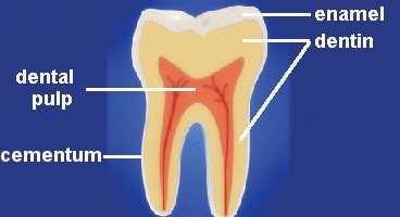
Each tooth consists of four parts:
- Enamel: It is the hardest substance in the body. Enamel is the hard, white outer covering of the tooth. It is mostly made of calcium phosphate, a rock-hard mineral.
- Dentine: A layer underlying the enamel. It is made of a hard calcified tissue. Dentine encloses the pulp cavity.
- Pulp: Pulp is a part of pulp cavity. It is a soft and gelatinous connective tissue. Blood vessels and nerves run through the pulp of the teeth.
- Cementum: A layer of connective tissue that binds the roots of the teeth firmly to the gums and jawbone.


Types of teeth

The teeth are classified into different types on the basis of its use. They are mainly used for cutting, biting, tearing, chewing and grinding.
They are differently shaped permanent teeth to break the food into small chunks which make the digestion easy.
Permanent teeth are divided into four types. Each has special structure and functions. They are:
They are differently shaped permanent teeth to break the food into small chunks which make the digestion easy.
Permanent teeth are divided into four types. Each has special structure and functions. They are:
- Incisors
- Canine
- Premolars
- Molars
Incisors

Incisors are situated in the center of both- top and bottom jaw. They have the following features:
- There are 4 incisors in each jaw.
- They are thin, flat-bottom, narrow- edged and sharp in shape.
- Incisors are mainly adapted for cutting.
- Incisors help us to make the initial bite.
Structure of Kidney

Kidneys - There is a pair of kidneys which are dark-red, each with bean-shaped with a notch, the hilus is present on its inner side. The blood vessels, ureter enter or leave the kidney through hilus. Each kidney is about 5 cm wide and 3 cm thick. In an adult male, the weight of the kidney is 150 gms and in the adult female, it weighs about 135 gms. They are situated in the anterior part of the abdomen protected by the pair of floating ribs (last two ribs). The left kidney is slightly lower than the left kidney.
Each kidney is made up of three layers renal cortex, renal medulla and renal pelvis. The renal medulla has 15 to 16 conical structure called medullary pyramids. The renal medullary pyramid ends into a structure called renal papilla. Columns of Bertini is present between the medullary pyramids. The medullary pyramids are connected to minor calyces. the minor calyces lead to major calyces. The major calyces open into the renal pelvis. The renal pelvis leads to the ureter. A kidney has about 10 lakh structural and functional units called nephrons.
Each kidney is made up of three layers renal cortex, renal medulla and renal pelvis. The renal medulla has 15 to 16 conical structure called medullary pyramids. The renal medullary pyramid ends into a structure called renal papilla. Columns of Bertini is present between the medullary pyramids. The medullary pyramids are connected to minor calyces. the minor calyces lead to major calyces. The major calyces open into the renal pelvis. The renal pelvis leads to the ureter. A kidney has about 10 lakh structural and functional units called nephrons.
Respiration in plants
Just like animals plants also respire (take oxygen in and release carbon dioxide). They use oxygen for the breakdown of food and release carbon dioxide. It can be represented by an equation as
Glucose (food) + Oxygen ---------> Carbon dioxide + water + energy.
Glucose (food) + Oxygen ---------> Carbon dioxide + water + energy.
- The intake of oxygen and release of carbon dioxide is through different routes in different parts of the plants.
- In leaves and stem, exchange of gases takes place through stomata (guarded pores).
- In stem with bark, exchange of gases take place through the lenticels.
- Root cells take the air (through diffusion) in the soil spaces.
- The oxygen in the air is then utilized for breaking down glucose to release energy and carbon dioxide.
- Respiration in most plants occurs under dark conditions (no light).
Importance of transpiration
Transpiration helps a plant in the following ways:
- Cooling effect: It helps in cooling of the surfaces of leaves thereby protecting them from excessive heat.
- Effect on ascent of sap and mineral transport: It helps in maintaining the concentration of the sap inside the plant body.
Functions of nephron.
Filtration of blood takes place in the Bowman's capsule. Blood reaches the glomerulus through afferent arteriole of the renal artery. The amount of blood that enters the Bowman's capsule while passing through glomerulus is called filtrate. The filtrate contains glucose, amino acid, salts, and water. This filtrate passes through renal tubule where selective reabsorption takes place. The useful substances like glucose, amino acid, water etc are reabsorbed. The remaining filtrate is called urine which is collected in the collecting duct.
Structure of nephron
A nephron is a microscopic structural and functional unit of the kidney. It is made of a renal corpuscle and a renal tubule. The renal corpuscle consists of a network of capillaries called glomerulus and Bowman's capsule. The corpuscle and tubule both are connected. They are made of epithelial cells. The tubule has five parts, namely
- Proximal convoluted tubule which is connected to the Bowman's capsule
- The loop of Henle which has two parts, ascending loop of Henle and descending loop of Henle.
- Distal convoluted tubule.
- The collecting tubule and
- Collecting ducts.
Structure of nephron
Nephrons are structural and functional units of the kidney.
Structure of nephron consists of
Structure of nephron consists of
- Malphigian corpuscles - It comprises glomerulus and Bowman's capsule .
- Renal tubule - It comprises Proximal convoluted tubule(PCT), Loop of henle, Distal convoluted tubule (DCT) and collecting duct.
Functions of stomach
The stomach is an elastic bag which stores food. It has the following functions:
- Gastric juices are secreted by the gastric glands present on the walls of the stomach.
- Different digestive juices (Hydrochloric acid, water, and an enzyme called pepsin) are mixed with the bolus. Pepsin helps to break down protein into simpler compounds (peptones).
- Another enzyme called rennin is used to convert the milk protein (casein) to curd.
- The food is churned thoroughly into the stomach with the digestive juices. This pulpy structure is called as chyme.
- After 3-4 hours of digestive process in the stomach, the food moves to the small intestine.
Functions of the small Intestine
The small intestine is a coiled tube. It has the following features and functions:
- It is responsible for chemical digestion of the food (chyme) and absorption of nutrients.
- The first part of the small intestine is called the duodenum, which receives chyme from the stomach. It also receives secretions from the liver (bile) and pancreas (pancreatic juices). Bile helps in digesting fats. Pancreatic juices like Amylase helps in converting starch to maltose, Trypsin helps to convert proteins and peptones to peptides and Lipase is responsible for converting fats into fatty acids and glycerol.
- The jejunum is the second part of the small intestine. No digestion takes place here.
- The last part of the small intestine is called ileum. Semi-digested food is received by them.
- Intestinal glands present on the walls of the ileum secrete intestinal juices like maltase, erepsin, sucrase, and lactase.
- The digestive process is completed in ileum when peptides are converted to amino acids in presence of erepsin, Maltose is converted to glucose with the help of maltase, sucrose is converted to glucose and fructose in presence of sucrase, and end products glucose and galactose are obtained in presence of lactase.
- The process of digestion is completed here.
- The simpler compounds are absorbed by the walls of the small intestine. The wall contains tiny finger-like projections called villi. Villi increase the surface area for absorption.
- The villi absorb amino acids and glucose and pass them to the blood system.
Functions of the large intestine
The large intestine is about 1.5 meters long. It has the following functions:
- The caecum acts as the junction between small and large intestines.
- The large intestine is responsible for absorbing water from the undigested food.
- The rectum stores the semi-solid feces which is eliminated out of the body through the anus.
Bowman's capsule
Bowman's capsule - It is a double layered cup-shaped structure. Its lumen is continuous with the lumen of the renal tubule. It has two layers:
- The outer parietal layer - made of squamous cells.
- The inner visceral layer - surrounds the glomerulus and is composed of a special type of cells called podocytes.
Bile
- It is yellowish green watery fluid produced in the liver.
- The colour of the bile is due to bilirubin and biliverdin.
- Bile does not have any digestive enzymes but it creates an alkaline medium, which is essential for the action of pancreatic enzymes.
- Emulsification of fat (fat is broken into fat droplets) takes place with the help of the bile juice secreted by the live and stored in gall bladder.
Diuresis
- If ADH is reduced, there is increase in production of urine, this is called as diuresis.
- A person affected with diuresis, loses a great amount of water along with essential mineral salts through excessive urination.
- Substances that increase the formation of urine are called as diuretics. For example, liquid, diets, tea, coffee etc.
Small intestine

Structure:The small intestine has three sub-regions:
Villi are supplied with a network of capillaries and a large lymph vessel called the lacteal.
Mucosa epithelium has goblet cells. They secrete mucus which helps in lubrication. Mucosa also forms crypts in between the bases of villi in the intestine (crypts of Lieberkuhn).
Functions:
- Duodenum: C- shaped
- Jejunum: A long coiled middle portion
- Ileum: Highly coiled. It opens in the large intestine.
Villi are supplied with a network of capillaries and a large lymph vessel called the lacteal.
Mucosa epithelium has goblet cells. They secrete mucus which helps in lubrication. Mucosa also forms crypts in between the bases of villi in the intestine (crypts of Lieberkuhn).
Functions:
- It serves for both digestion and absorption.
- It receives two digestive juices; the bile and pancreatic juice.
- Ileum is very long, has large villi and made up of single epithelium, which helps in absorption of food.
Artificial kidney (hemodialysis)
- In case of kidney failure, an artificial device is used to remove the nitrogenous waste products from the blood. This process is known as hemodialysis.
- The patient's blood is slowly pumped from the patient's body into the dialyzer, where waste products and extra fluid are removed and the purified blood is then returned into the same vein.
Excretion in plants
Removal of the waste and toxic products from the body is called as excretion. All the living organisms excrete their wastes. Like animals, plants do not have a special organ for excretion. They excrete through their vegetative parts only.
- Oxygen is a by-product of photosynthesis, which is excreted from the leaves of plants.
- Excess water is excreted as water vapour by the process of transpiration.
- Plants store there some of the excretory products in leaves and get rid of that by losing them.
- Some of the plants store their excretory product as resins, gums, latex, oils etc in stems, leaves and bark. Eventually, plant shed off their parts and thus excretion occurs.
- Plant produces secondary metabolite that are not used by plants but used by animals such as alkaloids.
- Alkaloids are the nitrogenous compound stored in different parts of the plant. It is used as a medicine, sedatives and insecticides.
- Excretion in aquatic plant takes place through diffusion.
Need for respiration
Respiration facilitates the oxygen to the tissues of the body. It plays a very important role such as:
- It produces ATP (energy), which drives all endergonic (energy-requiring) processes of the body.
- Without any energy, the body's systems would shut down, and life would stop.
- It expels the toxic carbon dioxide.
Anaerobic respiration in animals
- Anaerobic means without air (an means without). Respiration in the absence of oxygen to produce the energy they require this is called as
anaerobic respiration. - Muscles need oxygen and glucose to respire aerobically and produce
the energy they require, these are carried to the muscle via the blood. - For vigorous exercise our heart and lungs would not be able to get sufficient oxygen to our muscles in order for them to respire. In this case muscles carry out anaerobic respiration.
- Glucose ----> Lactic acid + Energy
Difference between anaerobic respiration in plants and animals
| Anaerobic respiration in plants | Anaerobic respiration in animals |
| 1. Products of anaerobic respiration is ethanol and | Products of anaerobic respiration is lactic acid |
| 2. Release more heat energy | Release less heat energy |
| 3. Released causes foaming | No release, so no foaming |

No comments:
Post a Comment